Home
Uncategories
Anatomy Of Ribs And Sternum : The Thoracic Cage | Anatomy and Physiology I / It also includes some facts regarding pathophysiology in this region.
Anatomy Of Ribs And Sternum : The Thoracic Cage | Anatomy and Physiology I / It also includes some facts regarding pathophysiology in this region.
Anatomy Of Ribs And Sternum : The Thoracic Cage | Anatomy and Physiology I / It also includes some facts regarding pathophysiology in this region.. Interactive tutorials about the ribs and sternum bones, with labeled images and diagrams featuring the beautiful illustrations of getbodysmart. Diagnose and treat somatic dysfunctions of the clavicle, sternum, and ribs. Learn more about the skeletal system with quizzes and labelling exercises. Manubrium, body (gladiolus), and xiphoid process 1. Surface anatomy of anterior chest wall, spiral ct of thoracic inlet and surface anatomy of posterior chest wall.
Wondering what the sternum is? It lies on the anterior thoracic wall in the middle. They increase in length, curvature and amount of cartilage craniocaudally. The sternum is comprised of three distinctive portions: Therefore in an anatomical position, the posterior end is higher and nearer the median plane in relation to the anterior end.
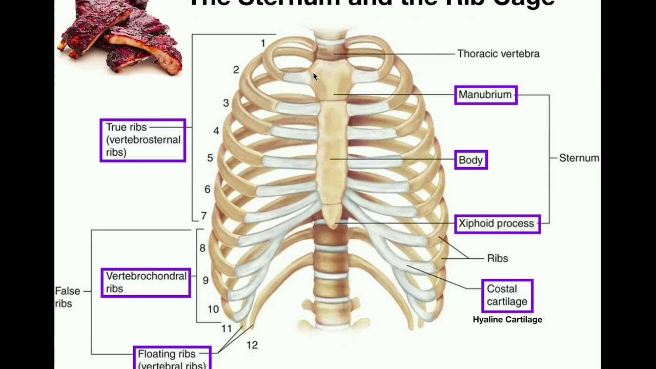
Anatomy | The Sternum, Rib Cage, & Vertebrae - YouTube from i.ytimg.com The number is the same in both males and females. The costotransverse ligaments in human: The chest wall is formed from the sternum anteriorly, 12 pairs of ribs, costal cartilages and intercostal muscles laterally, and the thoracic vertebrae posteriorly. The sternum is a flat, long bone that forms the medial and anterior part of the thoracic cage. The classification of human ribs. True ribs, false ribs, and floating ribs. Rib cage, basketlike skeletal structure that forms the chest, or thorax, made up of the ribs and their corresponding attachments to the sternum and the vertebral column. Sternum, costal cartilages, and ribs image source:
Each pair articulates with a.
Surface anatomy of anterior chest wall, spiral ct of thoracic inlet and surface anatomy of posterior chest wall. Describe the bony and cartilaginous articulations of the sternum and clavicle. It has clear front, side, and back planes. Ribs eight to ten are the false ribs and are connected to the sternum indirectly via the cartilage of the rib above them. There are twelve pairs of ribs. The first portion, the manubrium, articulates with both the clavicle and first rib and is therefore. Therefore in an anatomical position, the posterior end is higher and nearer the median plane in relation to the anterior end. Regional vertebrae (cervical, thoracic, lumbar), rib, sternum, os coxae, clavicle, scapula, humerus, ulna and radius for dr. Wondering what the sternum is? Attach the ribs to the costal cartilages. Interactive tutorials about the ribs and sternum bones, with labeled images and diagrams featuring the beautiful illustrations of getbodysmart. The ribs stretches posteriorly from thoracic vertebrae to the anterior lateral edges of the sternum. The thoracic cage (rib cage) is the skeletal framework of the thoracic wall, which encloses the thoracic cavity.
Sternum, costal cartilages, and ribs image source: Join us in this video where we show the sternum and rib articulation anatomy through the use of a model. Ribs are greatly reduced and never meet sternum each thoracic rib has two segments:. It consists of the ribs, the sternum, and the thoracic vertebrae, to which the ribs articulate. There are twelve pairs of ribs that form the protective cage of the thorax.
Sternum pain: Causes and when to see a doctor from i0.wp.com Ribs eight to ten are the false ribs and are connected to the sternum indirectly via the cartilage of the rib above them. There are two classifications of ribs. Anatomical variations of the sternum include varying sizes of the sternal angle. Attach the ribs to the costal cartilages. Sternum, costal cartilages, and ribs image source: Anterior view of the thoracic cage image source. It consists of the ribs, the sternum, and the thoracic vertebrae, to which the ribs articulate. The sternum or breastbone is a long flat bone located in the central part of the chest.
Therefore in an anatomical position, the posterior end is higher and nearer the median plane in relation to the anterior end.
It connects to the ribs via cartilage and forms the front of the rib cage, thus helping to protect the heart, lungs, and major blood vessels from injury. The thoracic cage consists of the 12 thoracic vertebrae, the associated intervertebral discs, 12 pairs of ribs with their costal cartilages, and the sternum. This guide gives a general overview of the anatomy of the thoracic spine. The sternum, commonly known as the breastbone, is a long, narrow flat bone that serves as the keystone of the rib cage and stabilizes the thoracic skeleton. These two bars fuse with each other along the middle line to form the cartilaginous sternum which is ossified. This often has little impact on function or treatment following injury but can however, cartilaginous connectors between the sternum and each of the upper six ribs assist with minor motions that occur with each breath. We examined the thoracic vertebrae last lab, so here we will only examine the ribs and sternum. Learn about sternum thorax ribs anatomy with free interactive flashcards. Ribs are greatly reduced and never meet sternum each thoracic rib has two segments:. The chest wall is formed from the sternum anteriorly, 12 pairs of ribs, costal cartilages and intercostal muscles laterally, and the thoracic vertebrae posteriorly. Moore'sanatomy and osteology human anatomy vertebra bones of the upper limb how to classified ribs and their shapes and fully describe human anatomy of upper parts. Lessons on the bone markings of the ribs and sternum. Learn all about this bone using our interactive anatomy image and detailed descriptions of its parts and function!
The length of ribs increases from 1st to 7th rib and after that slowly falls; Construct a robo skelly rib cage and the pelvis using the bucket method. Regional vertebrae (cervical, thoracic, lumbar), rib, sternum, os coxae, clavicle, scapula, humerus, ulna and radius for dr. Diagnose and treat somatic dysfunctions of the clavicle, sternum, and ribs. This is an online quiz called ribs and sternum anatomy.
Anatomy Of The Rib Cage Diagram from www.anatomynote.com The chest wall is formed from the sternum anteriorly, 12 pairs of ribs, costal cartilages and intercostal muscles laterally, and the thoracic vertebrae posteriorly. There are twelve pairs of ribs. The length of ribs increases from 1st to 7th rib and after that slowly falls; Surface anatomy of anterior chest wall, spiral ct of thoracic inlet and surface anatomy of posterior chest wall. Costae are arranged in pairs and articulate with two successive vertebrae. Moore'sanatomy and osteology human anatomy vertebra bones of the upper limb how to classified ribs and their shapes and fully describe human anatomy of upper parts. It discusses the specific anatomy of the ribs and costal cartilages, along with the sternum. Individual ribs have a bony dorsal part, a body of rib, and ventral costal cartilage.
The chest wall is formed from the sternum anteriorly, 12 pairs of ribs, costal cartilages and intercostal muscles laterally, and the thoracic vertebrae posteriorly.
The thoracic cage consists of the 12 thoracic vertebrae, the associated intervertebral discs, 12 pairs of ribs with their costal cartilages, and the sternum. The number is the same in both males and females. Each pair articulates with a. Anterior view of the thoracic cage image source. There are twelve pairs of ribs. Lessons on the bone markings of the ribs and sternum. Describe the bony and cartilaginous articulations of the sternum and clavicle. The thoracic cage (rib cage) is the skeletal framework of the thoracic wall, which encloses the thoracic cavity. Learn more about the skeletal system with quizzes and labelling exercises. The costotransverse ligaments in human: It has clear front, side, and back planes. The first portion, the manubrium, articulates with both the clavicle and first rib and is therefore. It lies on the anterior thoracic wall in the middle.
Diagnose and treat somatic dysfunctions of the clavicle, sternum, and ribs anatomy of ribs. Interactive tutorials about the ribs and sternum bones, with labeled images and diagrams featuring the beautiful illustrations of getbodysmart.


0 Comments:
Posting Komentar