Back Of Neck Anatomy : Human Anatomy for the Artist: May 2013 : The sections below will cover these elements in more detail.. Clinically, surface anatomy is used to split the neck. The neck is a complex anatomic region between the head and the body. This article covers the anatomy of the deep muscles of the back, including their function, blood supply, innervation, origin and insertion. Instant anatomy is a specialised web site for you to learn all about human anatomy of the body with diagrams, podcasts and revision questions. They control the scapulae (shoulder blades), which play a role in shrugging, neck movement, head support.
The spine runs from the base of your skull down the length of your back, going all the way down to your pelvis. This mri neck axial cross sectional anatomy tool is absolutely free to use. Watch cervical muscle anatomy animation. Bones of the neck picture. Clinically, surface anatomy is used to split the neck.
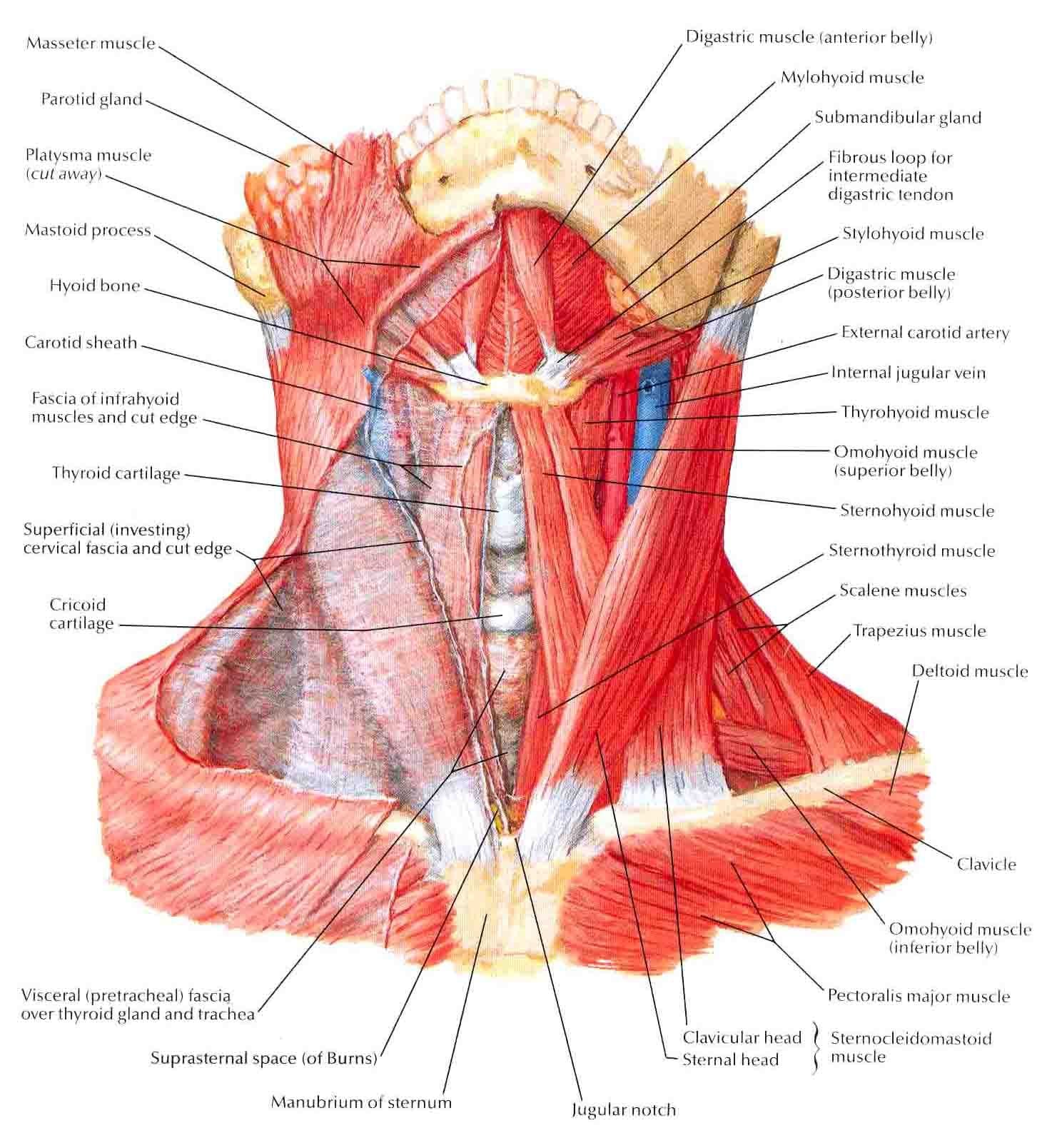
somso+arm+muscle+model+labeled | A&P.2.Skin.Bone.Muscle ... from s-media-cache-ak0.pinimg.com « back show on map ». This article describes the anatomy of the head and neck of the human body, including the brain, bones, muscles, blood vessels, nerves, glands, nose, mouth, teeth, tongue, and throat. Learn everything about the neck anatomy with this topic page. Discography is a diagnostic procedure the back experts at the southeastern spine institute (ssi) use to determine if any of your intervertebral discs are the primary cause of your back pain. The anatomy of the head and neck is complex because so many different functional structures are located close to each other. When most people mention their back, what they are actually referring to is their spine. Surface anatomy and surface markings bibliographic record list of illustrations subject index. In the back of the neck fascia envelops the trapezius muscle, extending to the occiput and upper nuchal line.
The neck is the area between the skull base and the clavicles.
The large spinous process (bump in back of neck) at c7 is called the vertebra prominens. Cervical spine anatomy is quite complex. Over the jugular notch presternum formed episternal in fig. Splenius capitis and cervicis are strong muscles that assist with major head and neck movements. Neck, in land vertebrates, the portion of the body joining the head to the shoulders and chest. C7 is the transition with the lumbar vertebrae and has many ligaments, and thus has a larger they have additional articular facets for the ribs. Attachment points for the muscles of the head and neck are located on the exterior surfaces of the skull and allow for important movement like. I love netter's anatomy books. The anterior jugular vein (v. This mri neck axial cross sectional anatomy tool is absolutely free to use. Want to learn more about it? Please review the anatomic diagrams on the following pages for details on organ structures and synonyms for organ names. Learn about these muscles, their locations & functional anatomy.
Please review the anatomic diagrams on the following pages for details on organ structures and synonyms for organ names. This article describes the anatomy of the head and neck of the human body, including the brain, bones, muscles, blood vessels, nerves, glands, nose, mouth, teeth, tongue, and throat. Want to learn more about it? They control the scapulae (shoulder blades), which play a role in shrugging, neck movement, head support. The diverse assortment of structures in the neck is naturally compartmentalised by a series of fasciae.
Neck Anatomy I (Netter's) - REBEL EM - Emergency Medicine Blog from rebelem.com Bones of the neck picture. At the back of the vertebral body are bony arches that project outward to form the facet joints and spinous processes. Attachment points for the muscles of the head and neck are located on the exterior surfaces of the skull and allow for important movement like. Discography is a diagnostic procedure the back experts at the southeastern spine institute (ssi) use to determine if any of your intervertebral discs are the primary cause of your back pain. It runs down the back part of the neck, and opens into the external jugular vein just below the middle of its course. The two costal articulation at the head and neck of ribs are both synovial joints. The neck is the area between the skull base and the clavicles. Use the mouse scroll wheel to move the images up and down alternatively use the tiny arrows (>>) on both side of the image to move the images.
Please review the anatomic diagrams on the following pages for details on organ structures and synonyms for organ names.
Navigate through the head and neck by the by type of body part you are looking for. Join our newsletter and receive our free ebook: At the back of the vertebral body are bony arches that project outward to form the facet joints and spinous processes. How many moveable vertebrae are in the… what are the main purpose of transverse… Over the jugular notch presternum formed episternal in fig. The large spinous process (bump in back of neck) at c7 is called the vertebra prominens. In the back of the neck fascia envelops the trapezius muscle, extending to the occiput and upper nuchal line. It arises from the inferior mental spine on the back of the symphysis menti and runs backward and slightly downward to be inserted into the anterior surface of the body of the. These bony elements naturally create a hollow opening in the center of the cervical spinal column—a canal that houses and. Jugularis anterior) begins near the hyoid bone by the. « back show on map ». The neck contains seven of these, known as the cervical vertebrae. The sections below will cover these elements in more detail.
The geniohyoid muscle is a narrow muscle, situated above the medial border of the mylohyoideus. Clinically, surface anatomy is used to split the neck. Some important structures contained in or passing through the neck include the seven cervical vertebrae and enclosed spinal cord, the jugular veins and carotid arteries, part of the esophagus, the larynx. Posterior cervical laminectomies or discectomies, as well as suboccipital. Cervical spine anatomy is quite complex.
Image result for neck muscles | Neck muscle anatomy ... from i.pinimg.com The back anatomy includes the latissimus dorsi, trapezius, erector spinae, rhomboid, & teres major. Our engaging videos, interactive quizzes. Neck, in land vertebrates, the portion of the body joining the head to the shoulders and chest. At the back of the vertebral body are bony arches that project outward to form the facet joints and spinous processes. Over the jugular notch presternum formed episternal in fig. The geniohyoid muscle is a narrow muscle, situated above the medial border of the mylohyoideus. Clinically, surface anatomy is used to split the neck. They control the scapulae (shoulder blades), which play a role in shrugging, neck movement, head support.
The neck is the start of the spinal column and spinal cord.
At the back of the vertebral body are bony arches that project outward to form the facet joints and spinous processes. The back comprises the spine and spinal nerves, as well as several different muscle groups. Learn about these muscles, their locations & functional anatomy. The neck is a complex anatomic region between the head and the body. Some important structures contained in or passing through the neck include the seven cervical vertebrae and enclosed spinal cord, the jugular veins and carotid arteries, part of the esophagus, the larynx. Posterior cervical laminectomies or discectomies, as well as suboccipital. The anterior jugular vein (v. See more ideas about anatomy, anatomy and physiology, muscle anatomy. The geniohyoid muscle is a narrow muscle, situated above the medial border of the mylohyoideus. Instant anatomy is a specialised web site for you to learn all about human anatomy of the body with diagrams, podcasts and revision questions. Muscles of the posterior neck and the back. The two costal articulation at the head and neck of ribs are both synovial joints. The diverse assortment of structures in the neck is naturally compartmentalised by a series of fasciae.


0 Comments:
Posting Komentar