Home
Uncategories
Leg Muscles Diagram Posterior - 11. Muscles of the Leg and Foot | Musculoskeletal Key - However, many of the leg muscles hip adductor muscles' attachment points.
Leg Muscles Diagram Posterior - 11. Muscles of the Leg and Foot | Musculoskeletal Key - However, many of the leg muscles hip adductor muscles' attachment points.
Leg Muscles Diagram Posterior - 11. Muscles of the Leg and Foot | Musculoskeletal Key - However, many of the leg muscles hip adductor muscles' attachment points.. Leg muscles diagram muscle diagram. Posterior muscles in the body. Lateral view of a pair of legs. It could be due to soft tissue injury. Those of the superficial group constitute a powerful muscular mass, forming the calf of the leg.
Leg muscles diagram muscle diagram. This muscle diagram is interactive: Muscles diagram front and back below you'll find several different muscles diagrams. Muscles that move the leg. The leg is separated into anterior, lateral, superficial posterior and deep posterior compartments by intermuscular septa and surrounded by the deep fascia of the leg.
Muscles of the Leg and Foot - Classic Human Anatomy in ... from schoolbag.info The posterior crural muscles—the muscles of the back of the leg are subdivided into two groups—superficial and deep. Knee muscles, posterior leg muscles anatomy, posterior thigh muscles. Lateral view of a pair of legs. Leg posterior 3d illustration project. It could be due to soft tissue injury. Start studying leg muscles (posterior view). The sacrum bone is almost always noticeable, no matter what the body type, because it is not covered with muscles or substantial fatty tissue. Muscles, connected to bones or internal organs and blood vessels, are in charge for movement.
Its action causes plantar flexion and inversion of.
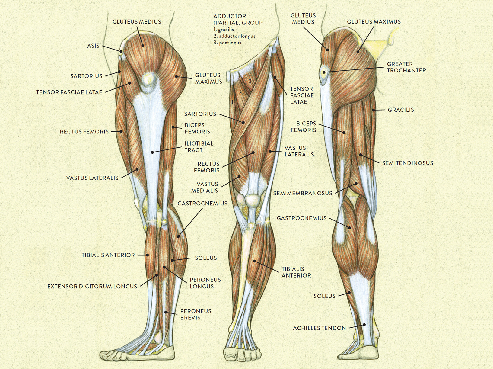
Muscles that move the leg. Leg muscle anatomy posterior leg muscles diagram photo album 10 / 10 ( 2 votes ) in this image, you will find tensor fascia latae, rectus femoris, vastus lateralis, iliopsoas, pectineus, adductor longus, gracilis, sartorius, vastus medialis, gluteus maximus, adductor magnus, semitendinosus, gracilis. Human leg muscles diagram leg muscle chart gosutalentrankco. The superficial muscles form the characteristic 'calf' shape of the posterior leg. Right posterior basal segmental bronchus. Male muscular system, full anatomical body diagram with muscle scheme, vector illustration educational poster. The posterior compartment of the leg is one of the fascial compartments of the leg and is divided further into deep and superficial compartments. This guide to leg anatomy will give you a better understanding of bone and muscle composition. This is the largest of the three compartments of the thigh. Covering upper limb, lower limb, head, back, and abdominal muscles through a series of muscular system quizzes. Medially rotates leg when flexed. The lower leg muscles are essential bodily structures. They all insert into the calcaneus of the foot (the heel bone), via the calcaneal tendon.
Some are small in length, and others are thinner and less bulky than muscles that extend or flex the knee or foot. Muscles that move the leg. It could be due to soft tissue injury. Human leg muscles diagram leg muscle chart gosutalentrankco. The posterior compartment of the leg is one of the fascial compartments of the leg and is divided further into deep and superficial compartments.
upper leg muscles common names Archives - Anatomy Body ... from i.pinimg.com This muscle diagram is interactive: Anatomy muscle 3d illustration 3d rendering adductor magnus anatomical arthritis back biceps femoris body buttocks calf muscle diagram female fitness gastrocnemius glutes gluteus maximus gracilis. Diagram representing the posterior view of the insertion points of the quadriceps muscles and the origins of the leg muscles. Those of the superficial group constitute a powerful muscular mass, forming the calf of the leg. Almost every movement in the body is the outcome of muscle contraction. 5 photos of the posterior leg muscles diagram. Some are small in length, and others are thinner and less bulky than muscles that extend or flex the knee or foot. The deep posterior compartment lies deep within the back of the lower leg.
The posterior crural muscles—the muscles of the back of the leg are subdivided into two groups—superficial and deep.
Have a product modelling and rendering project?. Male muscular system, full anatomical body diagram with muscle scheme, vector illustration educational poster. Its action causes plantar flexion and inversion of. Start studying leg muscles (posterior view). Muscles within this compartment primarily produce ankle plantarflexion as all 3 muscles form the achilles tendon. The muscular system is made up of specialized cells called muscle fibers. Leg posterior 3d illustration project. The superficial muscles form the characteristic 'calf' shape of the posterior leg. The following diagram illustrates the actions of the terms adduction, abduction, flexion anterior compartment thigh muscles. 5 photos of the posterior leg muscles diagram. The deep posterior compartment lies deep within the back of the lower leg. Flexion, medial rotation and adduction at hip. Those of the superficial group constitute a powerful muscular mass, forming the calf of the leg.
Lateral view of a pair of legs. The deep muscles that impact leg movement are generally smaller that those that are directly involved in flexing the knee. This is the largest of the three compartments of the thigh. The sacrum bone is almost always noticeable, no matter what the body type, because it is not covered with muscles or substantial fatty tissue. Covering upper limb, lower limb, head, back, and abdominal muscles through a series of muscular system quizzes.
Trumpa treniruotė stipresnėms kojoms | Tėtis, sportas ir ... from teciosportas.files.wordpress.com Muscles within this compartment primarily produce ankle plantarflexion as all 3 muscles form the achilles tendon. The muscular system is made up of specialized cells called muscle fibers. The deep muscles that impact leg movement are generally smaller that those that are directly involved in flexing the knee. The sacrum bone is almost always noticeable, no matter what the body type, because it is not covered with muscles or substantial fatty tissue. Lateral view of a pair of legs. Human leg muscles diagram leg muscle chart gosutalentrankco. Dissection of right lateral cervical region diagram. This is the largest of the three compartments of the thigh.
Muscles that move the leg.
Start studying leg muscles (posterior view). The posterior compartment of the leg is supplied by the tibial nerve. The sacrum bone is almost always noticeable, no matter what the body type, because it is not covered with muscles or substantial fatty tissue. Anatomy muscle 3d illustration 3d rendering adductor magnus anatomical arthritis back biceps femoris body buttocks calf muscle diagram female fitness gastrocnemius glutes gluteus maximus gracilis. Get in touch with us today! The superficial muscles form the characteristic 'calf' shape of the posterior leg. Rotator cuff muscle with anatomical posterior and anterior view expample. Right posterior basal segmental bronchus. The muscle of the anterior compartment (arm in anatomical position) function as flexors while the muscles of the posterior compartment function as extensors. Dissection of right lateral cervical region diagram. Their large size is one of the most characteristic features of the muscular. Female hip and leg muscles labeled posterior view, 3d rendering. Two muscles in the back of the leg pull on the achilles tendon as shown in figure 5.
This muscle diagram is interactive: leg muscles diagram. What total force do they exert?


0 Comments:
Posting Komentar